- Our Services
- Industries
- Automotive Materials and Components
- Electronic and electrical
- Ship
- Industrial detection
- Environmental detection
- Service
- Test
- Foreign Material Analysis-Formulation Analysis
- Authentication
- Train
- Failure analysis
- Laboratory Technical Services
- Judicial expertise
- Insurance technology appraisal
- Price evaluation
-

Our Services
Upgrade the quality level of Made in China
-
Resource Center
-

Test application form
For testing requirements, please download the test application form
-

FALAB People
Falab people become what you want!
-
-
News
-

Company News
Get the latest business news
-

Industry Information
Deliver the latest information and analyze the industry hot spots
-
-
Join Us
-

Recruitment
Falab, welcome to join us.
-
-
About FALAB
-
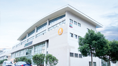
FALAB Introduction
China's third party testing and certification services pioneer and leader
-

Culture
To contribute to the quality and technology improvement of China's manufacturing industry
-